Optos
Optomap retinal imaging uses a special camera to obtain a wide-field view of the internal structures of the eye:
- high definition images
- view 80% of the retina at one time
- helps to identify peripheral retinal disease
- early diagnosis of retinal holes and tears
Corneal Topography
Corneal topography is performed with a non-contact instrument called a corneal topographer. These instruments work by projecting light rings onto the cornea and then using a computer to analyze the reflected image. Many corneal conditions can produce corneal shape anomalies such as irregular astigmatism that result in visual impairment.
Retinal Scanning Lasers
Retinal scanning lasers are non-contact instruments that utilize low-power laser energy to measure the thickness and physical features of the various layers of the retina.
Important clinical functions of retinal scanning lasers include the following:
- earlier and more accurate diagnosis of retinal disease
- assist in determining treatment plans
- preoperative surgical risk (cataract surgery, macular degeneration, diabetic retinopathy)
- postoperative surgical management

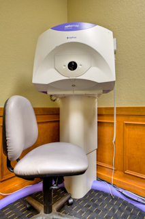
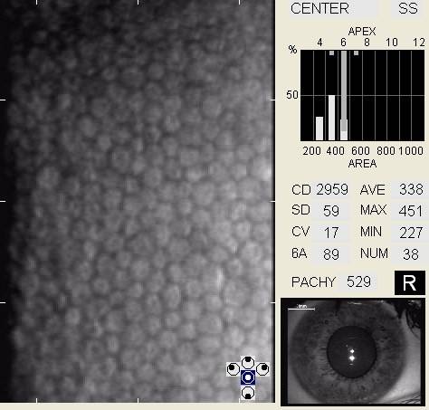
Specular Microscopy
To best way to perform specular microscopy is with a modern non-contact specular microscope.
The microscope captures an image of the endothelial cells on the back surface of the cornea and the doctor analyzes the results to make an assessment about the health of your cornea.
Specular microscopy may be indicated in the following clinical situations:
- contact lens wear
- evaluation of uveitis
- evaluaton of corneal edema
- evaluation of corneal opacity
- evaluation of neovascularization
- evaluation of endotheliopathies
- preoperative risk assessment (cataract surgery, glaucoma surgery, refractive surgery)
- postoperative surgical management
Visual Field Examination
Formal visual field examinations are generally performed using automated perimetry. These instruments work by measuring the ability of persons to see points of light at varying locations on a curved surface.
Visual field examinations may be reasonable and medically necessary in any of the following clinical circumstances:
- glaucoma
- glaucoma suspect
- other optic nerve disease
- neuro-ophthalmic disease
- retinal disease
- systemic disease
- vascular conditions affecting the visual fields
- high-risk medications affecting the visual fields
- disorders of the eyelids affecting the visual fields
- sigificant eye injury
- new functional limitations (can be reported by family members as “running into things”)
- unexplained vision loss (can be described as “trouble seeing” or “vision going in and out”)

Refraction
During an eye examination, your doctor may discover a vision problem like nearsightedness, farsightedness or astigmatism. If so, it’s likely that one of the next steps will involve a phoropter.
A phoropter is special machine used to switch multiple lenses in front of your eyes. It is used to measure the optical power of your eyes.
By having you look through the phoropter at a visual reference, the person performing the refraction can determine an ophthalmic lens prescription that produces clear, comfortable, binocular vision.

